
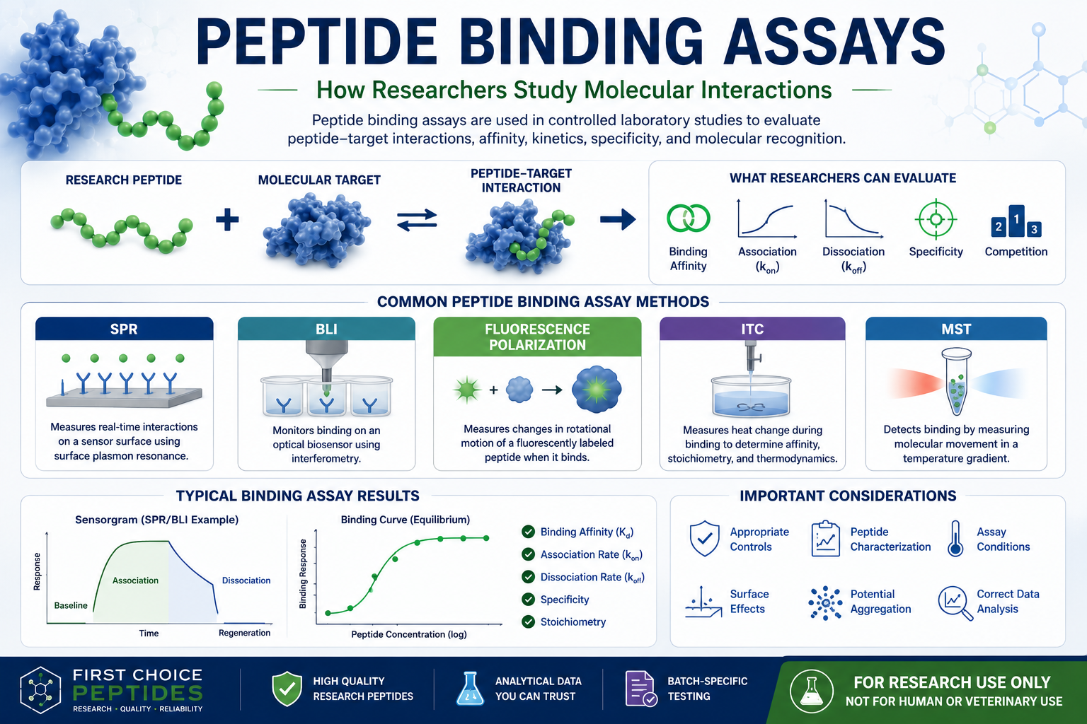
Learn how peptide binding assays are used in controlled laboratory research to evaluate peptide-protein interactions, binding affinity, specificity, association and dissociation kinetics, and molecular recognition.
Peptide Binding Assays: How Researchers Study Molecular Interactions
A peptide binding assay is a controlled laboratory experiment used to evaluate whether a peptide interacts with another molecular component under defined research conditions.
The other molecular component may be:
A purified protein
An enzyme
An antibody
A receptor preparation
Another peptide
A synthetic binding partner
A membrane model
Another defined laboratory target
Depending on the assay method and experimental design, researchers may investigate:
Whether measurable binding occurs
Relative or quantitative binding affinity
Binding specificity
Association and dissociation behavior
Competitive binding
Binding-site recognition
Interaction stoichiometry
Effects of peptide sequence changes
Effects of defined assay conditions
A binding result describes an interaction within the experimental system that was tested. It does not automatically establish a physiological, therapeutic, clinical, human, or veterinary effect.
Research-use notice: This article discusses peptide binding solely in the context of biochemical, biophysical, analytical, in-vitro, and controlled laboratory research. It does not provide information or instructions concerning administration, dosing, preparation for administration, diagnosis, prevention, treatment, clinical use, human use, or veterinary/animal use.
What Is a Peptide Binding Assay?
A peptide binding assay measures an interaction between a defined peptide and another molecular target.
A basic experimental model can be represented as:
Peptide + Molecular Target ⇌ Peptide–Target Complex
Researchers can monitor this interaction using an analytical signal generated by the selected assay.
Depending on the technique, the laboratory may measure:
Changes at an optical sensor surface
Interference patterns
Fluorescence behavior
Heat generated or absorbed during binding
Movement of molecules through a temperature gradient
Competition between labeled and unlabeled molecules
Another measurable analytical response
Different binding methods provide different types of information. No single assay automatically measures every characteristic of a peptide-target interaction.
What Are Peptide-Protein Interactions?
A peptide-protein interaction occurs when a peptide associates with a protein under specified experimental conditions.
The interaction can be influenced by molecular characteristics such as:
Peptide amino-acid sequence
Peptide length
Charge distribution
Hydrophobicity
Conformation
Chemical modifications
Protein structure
Accessible binding sites
Buffer composition
Temperature
pH
Ionic strength
Researchers may compare related peptide sequences to investigate which structural features contribute to measurable interaction with a protein.
For example, a laboratory could compare:
Original peptide sequence
with:
A sequence containing one amino-acid substitution
The resulting binding data may help researchers investigate whether that specific sequence position contributes to molecular recognition.
The observation applies to the experimental system and conditions tested.
What Can a Peptide Binding Study Reveal?
Depending on the method, a peptide binding study may help researchers evaluate several analytical characteristics.
Binding Detection
Does the assay produce a measurable signal consistent with an interaction between the peptide and molecular target?
Binding Affinity
How strongly do the two molecular partners associate under the defined assay conditions?
Binding Specificity
Does the peptide show greater measurable interaction with one target than with control or comparison targets?
Binding Kinetics
How quickly does the peptide associate with and dissociate from the target?
Binding Stoichiometry
How many peptide molecules appear to associate with each target molecule under the analytical model?
Competitive Binding
Can another molecule reduce or displace the measured peptide-target interaction?
The ability to determine each characteristic depends on the assay method, experimental design, data quality, and mathematical model.
What Is Peptide Binding Affinity?
Binding affinity describes the strength of a reversible molecular interaction under defined experimental conditions.
It is often represented using an equilibrium dissociation constant:
Kᵈ
The precise interpretation of Kᵈ depends on the binding model and assay.
Within an appropriate comparison using the same model and experimental conditions, a lower Kᵈ generally corresponds to tighter measured binding, while a higher Kᵈ generally corresponds to weaker measured binding.
Binding-affinity measurements are commonly generated by varying the concentration of one molecular partner and evaluating the resulting binding response. The reliability of the result depends on experimental design, concentration accuracy, model selection, and whether the assumptions of the model are satisfied. (PubMed Central (PMC))
What Does Kᵈ Mean?
Kᵈ is commonly called the:
Equilibrium dissociation constant
For a simple reversible interaction:
Peptide + Target ⇌ Complex
Kᵈ describes the relationship among the unbound peptide, unbound target, and peptide-target complex at equilibrium.
It is important not to treat a Kᵈ value as an inherent universal number that is completely independent of experimental conditions.
Measured affinity can be influenced by:
Assay format
Temperature
Buffer
pH
Ionic strength
Target presentation
Peptide labeling
Immobilization
Data-analysis model
Molecular concentration
Nonspecific interactions
Comparisons are strongest when the same method and closely matched experimental conditions are used.
What Are Association and Dissociation?
Peptide-target binding is often described as a dynamic process.
Association
Association occurs when the peptide and target come together to form a measurable complex.
The association-rate constant may be written as:
kₒₙ
or:
kₐ
Dissociation
Dissociation occurs when the peptide-target complex separates.
The dissociation-rate constant may be written as:
kₒff
or:
kᵈ
The lowercase kinetic symbol for the dissociation rate should not be confused with the uppercase equilibrium dissociation constant, Kᵈ.
Surface plasmon resonance methods can monitor association and dissociation in real time and can be used to derive kinetic and affinity information when the data fit an appropriate interaction model. (PubMed Central (PMC))
Binding Affinity vs. Binding Kinetics
Affinity and kinetics are related, but they are not identical descriptions.
Binding Affinity
Affinity describes the overall strength of the interaction at equilibrium.
Binding Kinetics
Kinetics describes how quickly the interaction forms and how quickly it separates.
Two peptide-target interactions could potentially have similar equilibrium affinity values while exhibiting different association and dissociation behavior.
For example:
One interaction may associate rapidly and dissociate rapidly.
Another may associate slowly and dissociate slowly.
Affinity alone does not provide the complete kinetic profile.
What Is a Direct Peptide Binding Assay?
In a direct binding assay, the experimental signal is generated by the peptide binding to the molecular target being studied.
A direct assay may involve:
Immobilizing the target and flowing peptide across it
Immobilizing a peptide and introducing the target
Labeling one molecular partner
Monitoring an interaction in solution
Measuring heat generated during direct binding
Direct assays can provide information about whether an interaction occurs and, depending on the method, its affinity or kinetics.
The way a molecule is attached, labeled, or presented may influence its behavior, so assay design must be considered during interpretation.
What Is a Competitive Peptide Binding Assay?
A competitive binding assay investigates whether one molecule competes with another for interaction with a molecular target.
A simplified design might include:
A labeled peptide or reference molecule
A molecular target
An unlabeled comparison peptide
Measurement of how the comparison peptide changes the reference signal
If increasing amounts of the comparison peptide reduce the reference binding signal, the data may be consistent with competition under the assay conditions.
Competition can indicate that two molecules interact with the same site, overlapping sites, or otherwise influence one another’s binding. The exact mechanism should not be assumed without supporting evidence.
Fluorescence-polarization methods have been used in controlled peptide competition and peptide-binding assays. (PubMed Central (PMC))
What Is a Saturation Binding Experiment?
A saturation experiment evaluates binding across a series of peptide concentrations.
As the peptide concentration increases, the measured interaction may increase until the available measurable binding capacity approaches a plateau.
A simplified curve may contain:
Low response at low concentration
Increasing response as concentration rises
A plateau as measurable sites become occupied
Appropriate mathematical analysis may be used to estimate affinity and binding capacity when the experimental model is valid.
A plateau alone should not automatically be interpreted as proof of specific binding. Controls are needed to investigate nonspecific signal and assay artifacts.
What Is Surface Plasmon Resonance?
Surface plasmon resonance, abbreviated SPR, is an optical biosensor method used to monitor molecular interactions at a sensor surface.
In a common SPR experiment:
One molecular partner is captured or immobilized on a sensor surface.
The other molecular partner flows across the surface.
Changes near the surface generate a response.
The response is monitored over time.
The resulting graph is commonly called a:
Sensorgram
SPR has been used to measure interactions involving synthetic peptides and proteins and can provide real-time association, dissociation, kinetic, and affinity information when appropriately designed. (PubMed Central (PMC))
How to Read an SPR Sensorgram
A typical sensorgram may contain several regions.
Baseline
The instrument records the signal before the peptide or target is introduced.
Association Phase
The analyte is introduced, and the response may rise as measurable complexes form.
Steady-State Region
Under some conditions, the response approaches a relatively stable level.
Dissociation Phase
The analyte is removed from the flowing solution, and the response may decrease as complexes dissociate.
Regeneration
In some assay designs, the sensor surface is treated to remove remaining bound material before another cycle.
Not every assay includes a regeneration step, and not every interaction follows a simple one-to-one binding model.
What Is Bio-Layer Interferometry?
Bio-layer interferometry, abbreviated BLI, is an optical method used to monitor molecular interactions at a biosensor surface.
A common BLI workflow includes:
Loading one molecular partner onto a biosensor
Establishing a baseline
Moving the sensor into a solution containing the other partner
Monitoring association
Moving the sensor into assay buffer
Monitoring dissociation
BLI can provide real-time kinetic and affinity information. Published laboratory protocols have used BLI to measure affinity and interaction kinetics between synthetic peptides and recombinant proteins. (PubMed Central (PMC))
SPR vs. BLI for Peptide Binding Research
SPR and BLI are both surface-based, label-free methods, but their instrument designs and workflows differ.
SPR
BLI
Monitors refractive changes near a sensor surface
Monitors optical interference changes at a biosensor
Commonly uses flow-based sample delivery
Commonly moves biosensors between sample wells
Can provide association and dissociation data
Can provide association and dissociation data
Can support kinetic and steady-state analysis
Can support kinetic and steady-state analysis
Requires attention to surface immobilization
Requires attention to biosensor loading
Neither method is automatically superior for every peptide interaction.
Method selection depends on:
Peptide and target properties
Available sample amount
Expected affinity range
Throughput requirements
Surface chemistry
Experimental objective
Instrument availability
What Is Fluorescence Polarization?
Fluorescence polarization, abbreviated FP, is a solution-based method that can be used to investigate molecular interactions.
A fluorescently labeled peptide rotates relatively quickly when it is free in solution.
When it binds to a larger molecular target, the complex generally rotates more slowly.
This change can alter the measured polarization signal.
Fluorescence-polarization methods can provide quantitative information about molecular interactions and have been used in peptide-binding and competition assays. (PubMed Central (PMC))
What Are the Advantages of Fluorescence-Polarization Assays?
Depending on the research system, FP may provide:
A solution-based assay format
No physical separation of free and bound fluorescent material
Compatibility with multiwell formats
Direct or competitive assay designs
Quantitative binding information
Relatively rapid measurements
However, the fluorescent label and its placement can influence peptide behavior.
Researchers should examine whether the labeled peptide still represents the interaction being investigated.
What Is Isothermal Titration Calorimetry?
Isothermal titration calorimetry, abbreviated ITC, measures heat generated or absorbed during a molecular interaction.
In a typical ITC study, one molecular partner is added incrementally to a solution containing the other partner.
The instrument measures heat associated with each addition.
Appropriate analysis can provide information concerning:
Binding affinity
Binding stoichiometry
Enthalpy
Entropy-related contributions
Free-energy relationships
ITC has been used to characterize peptide-protein binding and can evaluate unlabeled interacting partners, although reliable results require suitable sample concentration, buffer matching, and measurable heat change. (PubMed Central (PMC))
What Is Microscale Thermophoresis?
Microscale thermophoresis, abbreviated MST, evaluates the movement of molecules in a microscopic temperature gradient.
Molecular binding can change characteristics such as:
Size
Charge
Solvation behavior
Movement within the temperature gradient
Those changes can be used to generate binding curves and estimate affinity under an appropriate experimental design.
Published MST protocols have been used to determine molecular-binding affinity while minimizing or avoiding certain purification requirements in specific research systems. (PubMed Central (PMC))
Comparing Common Peptide Binding Assays
Method
Typical Information
Label or Surface Consideration
SPR
Association, dissociation, affinity, sensor response
One partner is generally attached or captured at a surface
BLI
Association, dissociation, affinity, sensor response
One partner is generally loaded onto a biosensor
Fluorescence Polarization
Solution-phase binding or competition
Usually requires a fluorescently labeled component
ITC
Affinity, stoichiometry, and thermodynamic information
No fluorescent label; requires measurable heat and sufficient sample
MST
Binding curve and affinity information
Often uses a fluorescent signal or intrinsic fluorescence
Pull-Down Assay
Evidence that components associate under assay conditions
Usually less directly quantitative
Plate-Based Binding Assay
Relative binding or competition signal
Surface attachment and detection reagents may affect interpretation
The appropriate method depends on the molecular system and the research question.
What Is Peptide Binding Specificity?
Binding specificity concerns whether a peptide shows measurable preference for one molecular target or site compared with alternatives.
Specificity can be investigated using:
Unrelated target controls
Sequence-altered peptides
Scrambled-sequence controls
Competition experiments
Target variants
Binding-site variants
Nonspecific surface controls
A measurable interaction with one target does not automatically establish specificity.
Specificity requires appropriate comparisons.
How Do Researchers Study the Effect of Peptide Sequence?
A peptide’s amino-acid sequence influences its molecular properties.
Researchers may compare:
Original sequence
Single-residue substitutions
Truncated sequences
Deletion variants
Modified terminal groups
Labeled and unlabeled versions
Scrambled-sequence controls
Differences in measured binding can help researchers investigate which sequence features contribute to the interaction.
These comparisons describe molecular behavior within the assay and should not be extrapolated automatically to human or veterinary outcomes.
Why Peptide Characterization Matters Before Binding Studies
A binding experiment is more meaningful when the material being studied has appropriate analytical documentation.
Researchers may review:
Peptide identity
HPLC purity
Molecular mass
Peptide content, when measured
Lot number
Certificate of Analysis
Stability information
Relevant modifications
If the peptide identity or composition is uncertain, interpretation of the binding result becomes more difficult.
Related guides:
[How Mass Spectrometry Supports Peptide Identity Testing]
[What Is HPLC Peptide Purity?]
[How to Read a Peptide Certificate of Analysis]
What Controls Are Important in Peptide Binding Assays?
Controls help researchers distinguish a meaningful interaction from background signal or assay artifacts.
Depending on the assay, useful controls may include:
Blank Control
Contains assay components without the peptide-target interaction being tested.
Target-Only Control
Contains the molecular target without the experimental peptide.
Peptide-Only Control
Contains the peptide without the intended target.
Nonspecific-Binding Control
Tests whether the peptide interacts with the assay surface, sensor, label, or unrelated material.
Negative-Control Peptide
Uses an unrelated or sequence-altered peptide expected not to show the same interaction.
Positive Control
Uses an established interaction when an appropriate reference is available.
Competition Control
Uses an unlabeled or comparison molecule to investigate whether a measured signal is competitively reduced.
The exact controls should match the assay design.
Why Concentration Accuracy Matters
Binding models depend on the concentrations of the interacting molecular partners.
An incorrect peptide concentration can affect:
Estimated affinity
Apparent stoichiometry
Curve shape
Comparison between experiments
Reproducibility
Researchers should distinguish between:
Total material weight
HPLC purity
Peptide content
Prepared assay concentration
These are not automatically interchangeable measurements.
For more detail, see:
[Peptide Purity vs. Peptide Content: What’s the Difference?]
Why Buffer Conditions Matter
Binding behavior can depend on the chemical environment.
Variables may include:
pH
Ionic strength
Salt composition
Detergents
Additives
Solvent percentage
Temperature
Reducing conditions
Metal ions
Buffer interactions
A peptide-target interaction measured under one set of conditions may differ under another.
Therefore, researchers should record and report the experimental conditions rather than treating affinity as entirely independent of assay context.
Can Peptide Labels Affect Binding?
Yes, potentially.
Peptides may be modified with:
Fluorescent labels
Biotin
Linkers
Affinity tags
Surface-capture groups
Other analytical modifications
A label may influence:
Charge
Molecular mass
Hydrophobicity
Steric accessibility
Conformation
Binding-site availability
Researchers should determine whether the labeled peptide remains an appropriate representation of the molecular interaction being studied.
Whenever possible, comparison with an unlabeled or alternatively configured assay can provide useful context.
Can Immobilization Affect Binding?
Surface-based methods generally require one binding partner to be attached or captured at a sensor surface.
Immobilization can potentially influence:
Molecular orientation
Accessibility of the binding site
Local surface density
Steric effects
Rebinding
Apparent kinetic behavior
Researchers should consider surface chemistry, loading level, reference surfaces, and assay orientation when interpreting surface-based results.
What Is Nonspecific Binding?
Nonspecific binding is assay signal arising from interactions other than the intended peptide-target interaction.
A peptide may interact with:
Sensor surfaces
Plates
Labels
Linkers
Blocking materials
Unrelated proteins
Aggregated material
Other assay components
Nonspecific signal can make binding appear stronger or more extensive than the intended molecular interaction.
Appropriate controls and reference subtraction help investigate this possibility.
What Is Peptide Aggregation in a Binding Assay?
Peptides can sometimes self-associate or form aggregates under particular experimental conditions.
Aggregation can affect:
Apparent concentration
Optical signals
Sensor responses
Nonspecific surface interactions
Binding-curve shape
Reproducibility
An unusual binding curve should not automatically be interpreted as evidence of a complex molecular mechanism.
Researchers may need to investigate material behavior, assay conditions, and data quality.
How to Read a Peptide Binding Curve
A simple equilibrium binding curve commonly displays:
X-axis: Peptide concentration
Y-axis: Measured binding response
Researchers may examine:
Whether the response rises with concentration
Whether the curve approaches a plateau
Whether replicate measurements agree
Whether controls show background signal
Whether the model fits the data
Whether the tested concentration range sufficiently defines the curve
A fitted value should not be interpreted without reviewing the underlying data and model assumptions.
How to Read Binding-Kinetics Data
Kinetic data may show:
Baseline
Association phase
Dissociation phase
Reference-subtracted response
Fitted kinetic model
Residuals between observed and fitted data
Researchers should consider:
Whether association and dissociation are adequately captured
Whether different concentrations produce consistent behavior
Whether the model fits the data
Whether mass-transport or rebinding effects may influence the result
Whether sensor loading is appropriate
Whether the controls are acceptable
A visually smooth fitted curve does not by itself prove that the selected binding model is correct.
What Does a Peptide Binding Assay Not Establish?
A peptide binding result does not automatically establish:
A functional response
A biochemical consequence beyond the measured interaction
Activity in a different laboratory system
Effects in humans
Effects in animals
Therapeutic efficacy
Clinical safety
Veterinary safety
Suitability for administration
Appropriate dosing
Peptide purity
Complete peptide identity
Absolute peptide content
Binding is one molecular characteristic measured under defined experimental conditions.
Separate questions require separate studies.
Binding Does Not Automatically Equal Function
A peptide may show measurable binding in an assay without producing a measurable downstream functional response.
Conversely, a complex experimental response may involve multiple interactions that are not fully explained by one direct binding measurement.
Researchers should distinguish among:
Binding
Biochemical activity
Cell-based assay response
and:
Other experimental outcomes
These are related research concepts, but they are not interchangeable.
Binding Data Should Not Be Extrapolated to Human or Veterinary Use
A peptide-target interaction observed in an in-vitro assay applies to the defined experimental system.
It does not automatically establish:
Absorption
Distribution
Metabolism
Stability in a living organism
Safety
Efficacy
Clinical outcome
Veterinary outcome
This article does not provide or imply authorization, suitability, or instructions for any human or veterinary application.
How to Evaluate a Peptide Binding Study
Researchers reviewing a peptide binding report can ask:
What peptide was tested?
Was the peptide analytically characterized?
Which target was used?
Which binding method was performed?
Was the assay direct or competitive?
Were appropriate controls included?
Which concentrations were evaluated?
Were replicates performed?
Were nonspecific signals investigated?
Which mathematical model was used?
Were affinity and kinetics distinguished?
Were the experimental conditions reported?
Does the conclusion remain within the limits of the assay?
Can the data be traced to the relevant peptide lot?
These questions provide more context than a single binding-affinity number.
Common Peptide Binding-Assay Mistakes
Mistake 1: Treating Binding as Proof of a Functional Effect
Binding and function are different experimental measurements.
Mistake 2: Comparing Kᵈ Values From Unrelated Assays Without Context
Different methods and conditions can produce different apparent affinity values.
Mistake 3: Ignoring Nonspecific Binding
Surface interaction, labeling, aggregation, or other assay components can create background signals.
Mistake 4: Assuming the Label Has No Effect
A fluorophore, linker, tag, or capture group can alter peptide behavior.
Mistake 5: Using an Inappropriate Binding Model
A mathematically fitted curve is only useful when the model represents the experimental system adequately.
Mistake 6: Confusing Affinity With Kinetics
The same equilibrium affinity can arise from different association and dissociation profiles.
Mistake 7: Ignoring Peptide Identity and Content
Uncertainty in the material being studied can compromise interpretation of the interaction data.
Mistake 8: Extrapolating Laboratory Binding to Human or Animal Outcomes
A molecular interaction in a controlled assay does not establish a clinical or veterinary result.
Frequently Asked Questions About Peptide Binding Assays
What is a peptide binding assay?
A peptide binding assay is a controlled laboratory experiment used to evaluate whether a peptide interacts with another molecular component under defined conditions.
What is a peptide-protein interaction?
It is a measurable association between a peptide and a protein in a laboratory research system.
What does binding affinity mean?
Binding affinity describes the strength of a reversible molecular interaction under the specified experimental conditions and analytical model.
What is Kᵈ?
Kᵈ is the equilibrium dissociation constant commonly used to describe binding affinity.
What is the difference between Kᵈ and kₒff?
Kᵈ describes equilibrium affinity. The lowercase kₒff describes the rate at which a measured complex dissociates.
What is the difference between affinity and kinetics?
Affinity describes the overall equilibrium interaction strength. Kinetics describes how quickly association and dissociation occur.
Can SPR measure peptide binding?
SPR can monitor molecular interaction at a sensor surface and can provide association, dissociation, kinetic, and affinity-related information when the assay is appropriately designed. (PubMed Central (PMC))
Can BLI measure peptide-protein interactions?
Yes. BLI has been used in laboratory protocols to measure peptide-protein interaction affinity and kinetics. (PubMed Central (PMC))
Can fluorescence polarization measure peptide binding?
Fluorescence-polarization assays can evaluate binding involving fluorescently labeled peptides and can support direct or competition formats. (PubMed Central (PMC))
What does ITC measure in a peptide binding study?
ITC measures heat associated with binding and can provide affinity, stoichiometry, and thermodynamic information under suitable experimental conditions. (PubMed Central (PMC))
Does peptide binding prove a therapeutic effect?
No. A binding result applies to the specific laboratory system tested and does not establish therapeutic efficacy, clinical safety, or suitability for human or veterinary use.
Peptide Binding Research at First Choice Peptides
At First Choice Peptides, our educational resources focus on analytical science, controlled laboratory research, peptide characterization, and research documentation.
Peptide binding data should be evaluated alongside information concerning:
Peptide identity
Chromatographic purity
Molecular mass
Peptide content, when measured
Batch traceability
Assay conditions
Experimental controls
Analytical method limitations
Continue learning:
[What Are Research Peptides Used For in Laboratory Studies?]
[How Mass Spectrometry Supports Peptide Identity Testing]
[What Is Mass Spectrometry in Peptide Testing?]
[HPLC vs. Mass Spectrometry for Peptide Analysis]
[Peptide Purity vs. Peptide Content: What’s the Difference?]
[Common Peptide Impurities: What Analytical Testing Can Detect]
[How to Read a Peptide Certificate of Analysis]
Final Takeaway
Peptide binding assays allow researchers to investigate molecular interactions under controlled laboratory conditions.
Depending on the method, a study may provide information about:
Binding detection
Affinity
Specificity
Association
Dissociation
Competition
Stoichiometry
Sequence-dependent differences
SPR, BLI, fluorescence polarization, ITC, MST, and other methods each provide different analytical capabilities.
The strongest interpretation considers:
The peptide
The target
The method
The controls
The experimental conditions
The data-analysis model
and:
The limits of the assay
Most importantly, peptide binding data should remain within the scope of the laboratory system tested.
A molecular binding observation does not establish therapeutic effects, clinical outcomes, or suitability for human or veterinary use.
Research use only