
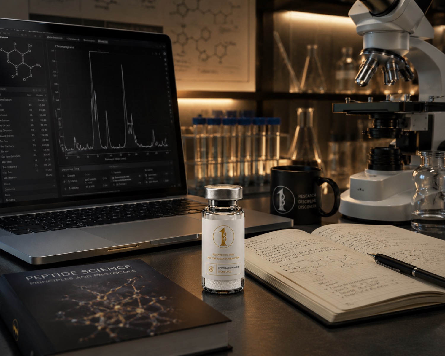
A model-by-model reading of the mouse and rat hair-follicle studies on thymosin beta-4, with no human data discussed anywhere. Research use only.
What the compound is
TB-500 is the acetylated heptapeptide Ac-LKKTETQ, residues 17 to 23 of thymosin beta-4, molecular formula C38H68N10O14. Thymosin beta-4 itself is the 43-residue parent polypeptide, and every follicle study cited below was carried out with thymosin beta-4 or with genetically modified rodents expressing or lacking it, not with the seven-residue fragment. All compounds discussed are supplied strictly as laboratory research materials. They are not for human or veterinary use, and nothing here is medical advice.
This article discusses no human data of any kind. There are no human studies described, summarised or implied below, and no statement here is about a person. Every finding named is a measurement taken in a mouse or rat model, and the model is named alongside the finding in each case.
The research question
Rodent skin is used to study follicle biology because the mouse hair cycle is synchronised and visible: in a mouse dorsal-skin preparation the follicle stage can be scored on histological section and the skin surface photographed on a fixed schedule, so a group difference can be quantified. The published question in this corpus is whether thymosin beta-4 levels or administration change those rodent follicle measurements, and through which cell population in the rodent follicle.
The endpoints across the mouse and rat studies are histological and cellular: follicle stage in mouse skin sections, follicle counts in mouse dorsal skin, migration and differentiation of follicle-derived cells isolated from rodent tissue, and transcript or protein levels measured in the same rodent tissue. No endpoint in this literature is a human endpoint.
It is worth stating how these rodent models are constructed, because the construction is what limits them. In a typical mouse dorsal-skin preparation the hair cycle is first synchronised across the animals in the group, so that follicles in mouse skin are at a comparable stage when the experiment begins. Measurements are then taken from the same mouse skin area on a fixed schedule, and the group difference is calculated within that mouse cohort. Nothing about that synchronised mouse preparation has a counterpart outside rodent models, which is exactly why the mouse is used for follicle work and exactly why a mouse follicle measurement stays inside the mouse model.
Mouse dorsal-skin and hair-cycle models
The most direct experiment in this group is a mouse study reporting that thymosin beta-4 induces mouse hair growth, in which the test system was mouse skin and the measurements were made on the mouse dorsal-skin surface and in mouse skin sections (PMID 26083021). Read strictly as a model description, the paper documents what was applied to mouse skin, over what window mouse skin was observed, and how follicle status in mouse tissue was scored.
A second study in this family examined the role of thymosin beta-4 in hair growth using rodent skin and molecular genetic methods, with transcript and protein measurements taken in the rodent tissue rather than a surface score alone (PMID 27130465). The value of that design in a mouse or rat preparation is that a tissue-level observation and a molecular measurement come from the same animals, so the two can be compared within the model rather than across separate rodent experiments.
Both of the mouse-skin studies above report tissue-level observations in mouse or rodent skin, and neither includes a human subject, human tissue or human endpoint of any kind. The measurements in those rodent studies are made on rodent skin sections and rodent skin surfaces, and the comparisons are between treated and control groups of the same rodent cohort. Any reading of those rodent results that reaches past the animal model is a reading the studies themselves do not support.
Rodent follicle stem-cell models
A separate line of rodent work asks which cell population inside the follicle changes. One study reported hair growth induced via stem cell migration and differentiation, using follicle-derived cells from rodent tissue as the assay system and cell migration and differentiation markers as the measured variables (PMID 17947589). The cells in that model are isolated from rodent follicles, and the readouts are cell-culture readouts taken from those rodent-derived cells.
An earlier report measured hair growth in a rodent model alongside activation of hair follicle stem cells in the same rodent tissue, combining a tissue-level score with a stem-cell marker measurement (PMID 14657002). Both studies are cell-and-tissue studies in rodents, and neither included any human subject or human tissue endpoint.
Group size is the plainest constraint on all of this rodent work. A mouse skin study typically compares small cohorts of animals, and a rodent follicle-cell experiment compares small numbers of culture replicates, so a difference measured in one mouse cohort can move considerably when the same mouse experiment is repeated. That is why the rodent findings above are described here as measurements taken in named mouse and rat models rather than as settled properties of the molecule, and why the review literature mapping those rodent models is treated as a map rather than as added evidence.
Angiogenesis and review literature in rodent systems
Follicle development in rodent skin is vascularised, and one study grouped angiogenesis, wound healing and hair follicle development as parallel endpoints measured in animal preparations of skin (PMID 15037013). Placing the three together in one rodent study is methodologically useful because vessel density and follicle status are scored on the same sections of animal tissue.
A later review collects the rodent follicle work and sets out the multiple roles proposed for thymosin beta-4 in follicle growth and development across those animal models (PMID 33393222). As a secondary source its role in a laboratory context is to map which rodent models exist and what each measured, not to add evidence of its own. No human model appears in that map, and none is introduced here.
The marker-based readouts in the rodent follicle studies deserve one further note. When a study reports stem cell activation in rodent follicle tissue, the underlying measurement is the abundance or localisation of specific marker proteins in that rodent tissue, detected by staining or immunoblot. A marker measurement in a rodent follicle preparation is an observation about that preparation, and the step from a rodent marker change to any statement about follicle biology in another species is a step none of the cited rodent studies took.
Limits of the evidence
The limits in this area are unusually strict and worth stating flatly. Every finding above was measured in mice, rats or cells isolated from rodent tissue. There is no human evidence discussed in this article, and no rodent follicle result described here transfers to a person, a human condition, or any cosmetic or medical question about people.
Beyond the species boundary, the rodent hair cycle is synchronised in a way that has no direct counterpart outside those animal models, which is precisely why the mouse is used and precisely why a mouse follicle measurement cannot be read across. Group sizes in the mouse and rat studies are small, most of the corpus tested the full-length parent polypeptide rather than the supplied fragment, and independent replication across unrelated laboratories is limited. These are preclinical rodent observations, and that is the whole of what they are.
Related materials in the catalogue
The product page is TB-500 and the compound guide is the TB-500 research guide, listed under tissue repair. The wound-model literature for the same parent polypeptide is covered in TB-500 and thymosin beta-4 in wound-model research, and skin-model work on a different material is covered in the GHK-Cu dermatology article.
Lot documentation is published under certificates, solvent volumes are worked out with the reconstitution calculator, and the analytical background is set out in how to verify a peptide certificate of analysis, HPLC peptide purity and mass spectrometry peptide testing. All compounds discussed are supplied strictly as laboratory research materials. They are not for human or veterinary use, and nothing here is medical advice.
References
- Thymosin Beta-4 Induces Mouse Hair Growth. PloS one, 2015. PMID 26083021
- Role of thymosin beta 4 in hair growth. Molecular genetics and genomics : MGG, 2016. PMID 27130465
- Thymosin beta 4 induces hair growth via stem cell migration and differentiation. Annals of the New York Academy of Sciences, 2007. PMID 17947589
- Thymosin beta4 promotes angiogenesis, wound healing, and hair follicle development. Mechanisms of ageing and development, 2004. PMID 15037013
- Multiple potential roles of thymosin β4 in the growth and development of hair follicles. Journal of cellular and molecular medicine, 2021. PMID 33393222
- Thymosin beta4 increases hair growth by activation of hair follicle stem cells. FASEB journal : official publication of the Federation of American Societies for Experimental Biology, 2004. PMID 14657002
Research use only