
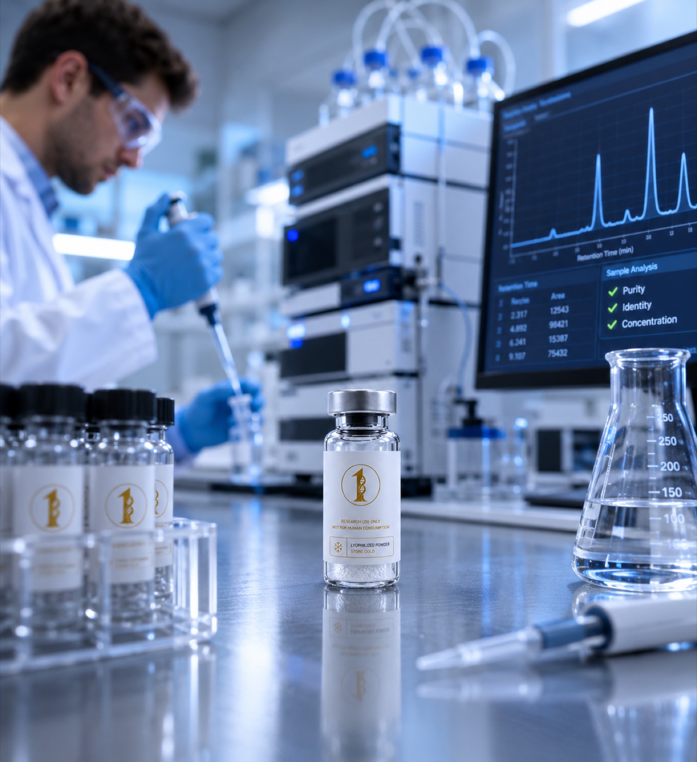
Cloudiness is light scattering from aggregates or undissolved solid. What drives it: pH, temperature, agitation, freeze-thaw and benzyl alcohol. Research use only.
Short answer
A clear peptide solution turns cloudy when particles large enough to scatter visible light appear in it. Those particles are almost always one of three things: undissolved solid that never went into solution, self-associated peptide aggregates that formed after dissolution, or precipitate thrown out because the solution conditions moved past the solubility limit of that sequence. pH, temperature, mechanical agitation, freeze-thaw cycling and the preservative in the diluent are the variables that decide which of the three occurs. A haze is a physical observation about the state of the material, and material in that state is no longer characterised, so it is not a valid input to an experiment. All compounds discussed are supplied strictly as laboratory research materials. They are not for human or veterinary use, and nothing here is medical advice.
What cloudiness physically is
Visible haze is Rayleigh and Mie scattering from particles in the low submicron to micron range. A monomeric peptide in solution is far too small to scatter appreciably, which is why a properly dissolved preparation looks like water. Turbidity therefore reports the appearance of new, larger species. The classic reference systems for this behaviour are insulin formulations, where physical stability has been studied for decades precisely because the visible change tracks a molecular one. A review of the physical stability of insulin formulations describes how a clear preparation converts to a turbid or fibrillar one and which formulation and handling variables govern that conversion (PMID 6341125).
The molecular event underneath the haze is usually self-association. Work on insulin aggregation in solution characterised how monomer associates into higher order species and how solution conditions and interfaces drive the process (PMID 2079389). The important consequence for a laboratory is that aggregation is generally not reversed by warming, shaking or waiting. The visible haze is the late stage of a process that began invisibly, so by the time it can be seen the soluble monomer concentration has already changed.
Cause one: it never dissolved
The most common source of a cloudy vial is not degradation at all. A lyophilized cake that has been hit with a fast jet of diluent, or one that was dried into a dense plug, can leave fine solid suspended rather than dissolved. Signs of incomplete dissolution are a haze that clears slowly on gentle standing, visible specks against a dark background, and a cake residue clinging to the vial wall or stopper.
Directing the diluent down the inside wall of the vial rather than onto the cake, then allowing several minutes of quiet standing with occasional gentle swirling, resolves most cases. Vortexing and vigorous shaking are counterproductive: they introduce air and create the air to liquid interface that promotes the second cause below. Peptides with strongly hydrophobic or highly aggregation-prone sequences may simply have a low aqueous solubility ceiling, in which case a lower concentration is the answer rather than more mechanical energy.
Cause two: aggregation after dissolution
A solution that was clear and later became hazy points at aggregation. Several triggers are well documented in the protein and peptide formulation literature.
pH near the isoelectric point
Net charge is what keeps peptide molecules electrostatically apart. When solution pH approaches the isoelectric point of the sequence, net charge falls toward zero, repulsion weakens and association becomes favourable. Solubility minima at the isoelectric point are a general property, and this is why the pH of the diluent, and any buffer or acid used to aid dissolution, is a variable that belongs in the notebook rather than an incidental detail.
Interfaces, agitation and shear
Air to liquid interfaces partially unfold peptide and protein molecules, exposing hydrophobic surface that then associates. Shaking a vial during transport, aggressive vortexing, and repeated draws that foam the solution all supply that interface. Studies of insulin physical stability identified agitation and surface contact as drivers of the transition from clear solution to turbid or fibrillar material (PMID 6341125, PMID 2079389).
Temperature and freeze-thaw
Warming increases conformational mobility and speeds association. Freezing does something less obvious: as ice forms, the peptide and the solutes concentrate in the shrinking unfrozen fraction, local pH can shift as buffer components crystallise at different rates, and a new ice to liquid interface is created. Each freeze and thaw cycle exposes the material to that sequence of events again, which is why a solution frozen and thawed repeatedly turns hazy sooner than one thawed once.
Concentration
Association is concentration dependent. A stock prepared at the top of the solubility range for that sequence can be clear at preparation temperature and cloudy after a night in the refrigerator, simply because solubility fell while concentration did not.
Cause three: the preservative
Bacteriostatic water contains benzyl alcohol as an antimicrobial preservative, and benzyl alcohol is not a passive spectator in a peptide or protein solution. Its effect has been characterised in several model systems. A mechanistic study of benzyl alcohol induced aggregation of recombinant human interleukin-1 receptor antagonist in aqueous solution set out how the preservative promotes association of that protein (PMID 15514986). A separate study of interferon alpha-2a examined the role of benzyl alcohol in unfolding and aggregation of that molecule (PMID 25100180). A broader investigation of alcohol induced protein aggregation established the general mechanism, namely that the alcohol stabilises a partially unfolded intermediate, and it is that intermediate rather than the native state that goes on to aggregate (PMID 20597088).
Two points follow for laboratory practice. First, preservative choice is an experimental variable: a solution prepared in preserved diluent and one prepared in unpreserved sterile water are not interchangeable samples of the same stock. Second, the trade is real in both directions. Unpreserved water gives no antimicrobial protection in a multi-draw vial, and microbial growth is itself a cause of turbidity, usually accompanied by a change in odour or a settling deposit over days. Single-use aliquots avoid needing to choose. Diluent specifications are described on the bacteriostatic water guide and the item itself is listed under bacteriostatic water.
Sequence features that raise the risk
Some catalogue materials carry structural features that make association more likely. TB-500 is the acetylated fragment Ac-LKKTETQ corresponding to residues 17 to 23 of thymosin beta-4, a short and highly soluble sequence. Tesamorelin, by contrast, is a 44-residue growth hormone releasing hormone analogue carrying an N-terminal trans-3-hexenoyl group, molecular formula C221H366N72O67S and molecular weight 5136.00 g/mol; a long chain with a lipophilic modification has more opportunity for hydrophobic self-association than a seven-residue fragment. CJC-1295 with DAC carries a maleimidopropionic acid lysine linker designed to bind serum albumin, which is another hydrophobic surface. Length, hydrophobic modification and net charge at working pH together predict which vials need the gentlest handling.
Practical laboratory handling
The following are handling practices for research material and carry no implication about use in a person.
- Add diluent slowly down the vial wall. Never squirt it directly onto the cake, and never vortex to force dissolution.
- Give it time. Several minutes of standing with occasional gentle inversion dissolves most cakes. Patience beats agitation.
- Inspect against light and dark backgrounds. Fine haze is easiest to see against black; larger particulate is easiest against white.
- Aliquot for single use. One thaw per tube eliminates freeze-thaw cycling, which is the most avoidable aggregation driver in routine work.
- Record the diluent. Preserved or unpreserved, lot, volume and date. Given the preservative effects above, this is data, not bookkeeping.
- Keep concentrations off the ceiling. Preparing below the solubility limit costs a little volume and removes a whole failure mode. Volumes and target concentrations can be calculated with the reconstitution calculator.
- Treat a hazy stock as uncharacterised. Its soluble concentration is no longer the label concentration, so it cannot serve as a quantitative input.
Confirming what the haze is
Appearance narrows the possibilities but does not identify the species. Chromatography separates monomer from covalently modified degradation products and shows whether the soluble fraction still matches the original trace, an approach described in the article on HPLC peptide purity. Mass measurement distinguishes an oxidised or hydrolysed molecule from an unchanged one that has merely self-associated, as covered in mass spectrometry peptide testing. The purity claim on the incoming lot is the baseline for both, and reading that document critically is covered in how to verify a peptide certificate of analysis, with lot documents published on the certificates page. If the incoming powder was within specification and the solution clouded afterward, the cause sits in preparation and storage rather than in the material as supplied.
All compounds discussed are supplied strictly as laboratory research materials. They are not for human or veterinary use, and nothing here is medical advice.
References
- Insulin aggregation in solution. International journal of peptide and protein research, 1990. PMID 2079389
- Physical stability of insulin formulations. Diabetes, 1983. PMID 6341125
- Mechanism for benzyl alcohol-induced aggregation of recombinant human interleukin-1 receptor antagonist in aqueous solution. Journal of pharmaceutical sciences, 2004. PMID 15514986
- Role of benzyl alcohol in the unfolding and aggregation of interferon α-2a. Journal of pharmaceutical sciences, 2015. PMID 25100180
- Role of partial protein unfolding in alcohol-induced protein aggregation. Proteins, 2010. PMID 20597088
Research use only