
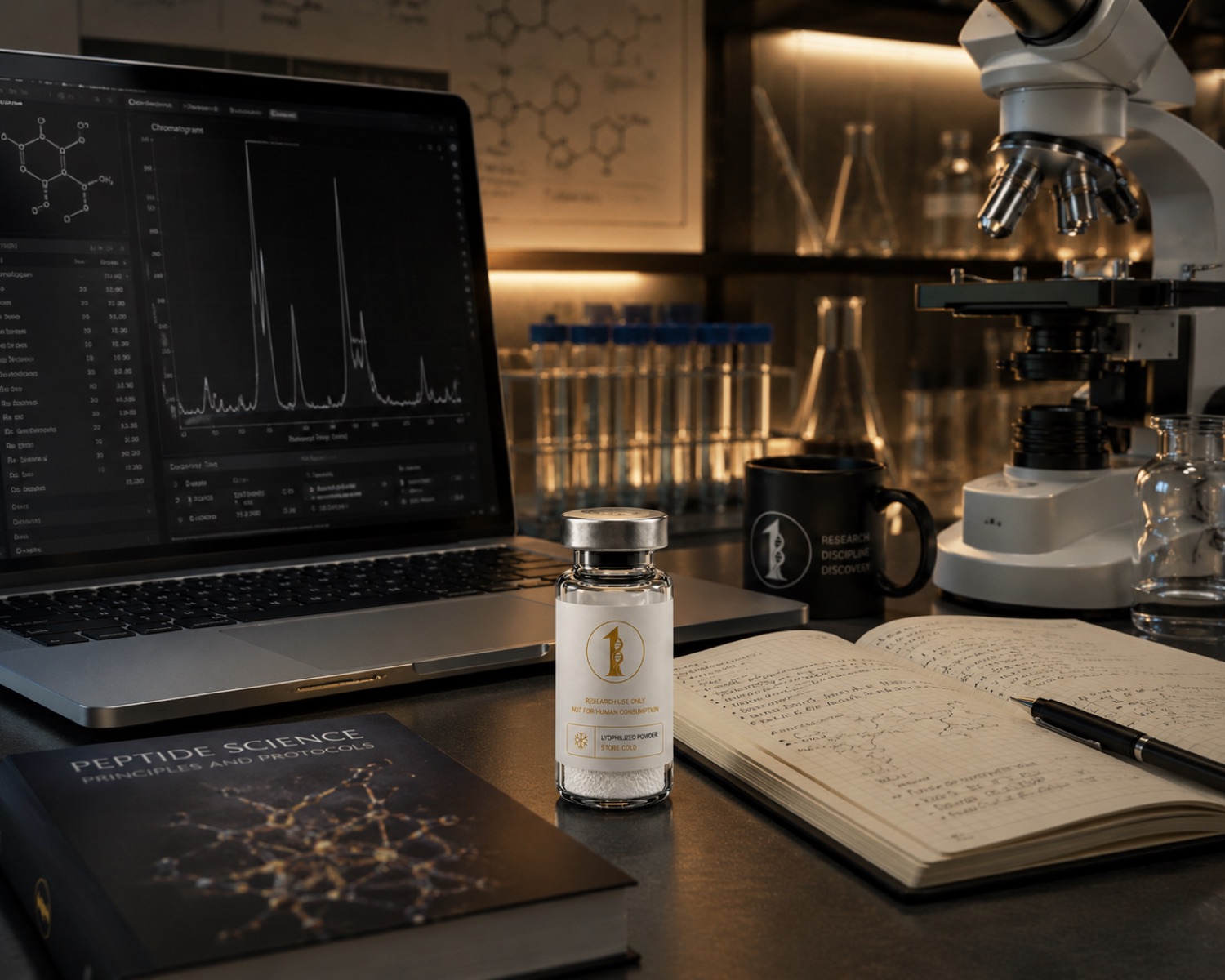
How preclinical studies examined BPC-157 in rat tendon, muscle, alkali-burn and gastric models, and what each experiment actually measured. Research use only.
What the compound is
BPC-157 is a synthetic pentadecapeptide of 15 amino acid residues, sequence GEPPPGKPADDAGLV, molecular formula C62H98N16O22, molecular weight 1419.50 g/mol, CAS number 137525-51-0. The published literature refers to it as a stable gastric pentadecapeptide, and review articles introduce it as a sequence characterised in gastric juice (PMID 34267654). In a research catalogue it is supplied as a lyophilised solid of stated purity with a certificate of analysis. All compounds discussed are supplied strictly as laboratory research materials. They are not for human or veterinary use, and nothing here is medical advice.
Because the peptide is short and unmodified, the identity checks that matter in a laboratory are the ordinary ones: an accurate mass consistent with the formula above, and a chromatographic purity figure measured against a documented method. Those checks are described separately in mass spectrometry peptide testing and HPLC peptide purity.
The sequence also sets the stability questions worth asking of stored material. There is no cysteine, so no disulfide chemistry has to be preserved, but the central region carries two adjacent aspartate residues, and aspartate motifs are the positions at which hydrolysis and isomerisation are most often examined in peptide stability work. Those are analytical questions about the supplied solid and its solutions, answered by chromatography on stored samples, and they are separate from anything in the biological literature summarised below.
The research question
The preclinical literature on BPC-157 asks a narrow experimental question. When a defined injury is created in a rodent tissue, or a defined stress is applied to a cultured cell population, what happens to the measured repair markers in the treated group compared with the control group. The endpoints are laboratory endpoints: outgrowth distance from a tendon explant, cell migration across a scratch, capillary density counted on a histological section, ulcer area in a stomach preparation, or receptor protein levels measured by immunoblot.
None of the studies summarised below were designed to answer a question about people, and none are read here as though they were. What the literature offers a laboratory is a set of named models, stated methods and measurable variables that another laboratory can repeat. Everything below is described in those terms and no further.
Tendon explant and tendon-healing models
The most frequently cited experimental work in this area is a rat tendon study that combined tissue explants with cultured cells (PMID 21030672). In that model, tendon tissue and isolated tendon fibroblasts were the test system, and the measured variables named in the report were tendon outgrowth from the explant, cell survival under culture stress and cell migration. Those three readouts are the practical reason the paper is repeated so often: outgrowth and migration can be quantified by image analysis, and survival by a standard viability assay, so the model is portable between laboratories.
A second rat study looked at muscle and tendon healing together and used angiogenesis as its comparative endpoint (PMID 20388964). Here the injury was surgical, the tissue was assessed histologically, and vessel counts were the variable placed alongside the healing measurement. Read as a laboratory design, the pairing is deliberate: a repair endpoint and a vascular endpoint measured in the same animals, so that changes in one can be examined against changes in the other rather than assumed.
A laboratory reading the explant study and the surgical study together will notice what the two designs do not share. The explant work removes cells from their tissue context and measures behaviour that can be imaged and quantified continuously, while the surgical work leaves the tissue intact and measures the result at fixed intervals after the injury. Neither substitutes for the other: one answers a question about cell behaviour under controlled culture conditions, the other a question about tissue at a defined point after a defined lesion. Where a review describes the two as agreeing, the agreement is an interpretation across different endpoints rather than a replication of the same measurement.
Angiogenesis, VEGFR2 and burn-wound models
A separate line of work asks what happens at the level of the endothelial receptor. In a study combining cell culture with animal experiments, vascular endothelial growth factor receptor 2 was the named target, and the measured variables were receptor activation and receptor up-regulation (PMID 27847966). Receptor phosphorylation and total receptor abundance are immunoblot endpoints, which places this paper in a different methodological family from the histology-based tendon work even though the tissue question overlaps.
An alkali-burn wound study in animals paired an in vivo wound model with in vitro assays of proliferation, migration and angiogenesis in cultured cells (PMID 25995620). The design is a common one in wound biology: the animal model provides the tissue-level measurement, and the cell assays isolate which cell behaviour changed. A review devoted specifically to the vascular literature collects the animal and cell studies in this family and describes the vessel-level observations reported across them (PMID 23782145).
Gastric and mucosal injury models
The oldest models in this literature are gastrointestinal. A rat gastric ulcer study measured lesion area and mucosal histology in treated and control animals (PMID 15052688), and that basic design, a chemically or physically induced lesion scored by area, is the template much of the later work follows in other tissues. A broad review of the wound-healing literature places the gastric models alongside the skin and tendon models and describes the range of animal preparations used across the field (PMID 34267654).
Two review articles extend the picture into mechanism-level questions that were investigated in rodents. One collects the experiments that examined the relationship between the peptide and the nitric oxide system in animal preparations, including the pharmacological blockade experiments those studies used (PMID 23755725). The other reviews rodent central nervous system models in which the peptide was administered and neurological or histological endpoints were recorded (PMID 34380875). Both are secondary sources, and in a laboratory context their usefulness is as a map of primary models rather than as evidence in themselves.
Limits of the evidence
Every study named above is preclinical. The subjects are rats, cultured rodent or human cells, or tissue explants, and the endpoints are laboratory measurements taken in those systems. Group sizes in the rodent work are small by the standards of clinical research, a large fraction of the corpus originates from a small number of collaborating groups, and independent replication across unrelated laboratories is thinner than the citation count suggests.
Publication practice in this field also favours positive findings, so the balance of reported results is not a balance of experiments performed. None of this makes the models uninteresting, but it does set the boundary of what can be said: these are observations in named animal and cell models, and they do not extrapolate to people. No human outcome is described in this article and none should be inferred from it.
The animal preparations also differ between reports in vehicle, formulation and delivery, so results are not straightforwardly poolable even where the tissue and the endpoint match. Vessel counting in particular is exposed to observer effects, since capillary density on a section depends on the stain, the fields chosen and whether the counter knew the group assignment, and reports vary in how fully those choices are documented. That is an ordinary situation in a preclinical literature of this age, and it is why a laboratory planning its own work reads these papers for their model descriptions rather than for their effect sizes.
Related materials in the catalogue
The compound page for this material is BPC-157, and the compound guide is the BPC-157 research guide. It sits in the tissue repair category alongside other materials used in repair-model work. Related mechanism articles cover TB-500 in wound-model research and the injury and recovery model literature as a whole.
For bench work, the reconstitution calculator converts a target concentration and vial mass into a solvent volume, and analytical documentation for each lot is published under certificates. Two method articles explain how to read that paperwork: how to verify a peptide certificate of analysis and what a 99 percent purity figure means. All compounds discussed are supplied strictly as laboratory research materials. They are not for human or veterinary use, and nothing here is medical advice.
References
- Stable Gastric Pentadecapeptide BPC 157 and Wound Healing. Frontiers in pharmacology, 2021. PMID 34267654
- The promoting effect of pentadecapeptide BPC 157 on tendon healing involves tendon outgrowth, cell survival, and cell migration. Journal of applied physiology (Bethesda, Md. : 1985), 2011. PMID 21030672
- BPC 157 and blood vessels. Current pharmaceutical design, 2014. PMID 23782145
- Therapeutic potential of pro-angiogenic BPC157 is associated with VEGFR2 activation and up-regulation. Journal of molecular medicine (Berlin, Germany), 2017. PMID 27847966
- Modulatory effect of gastric pentadecapeptide BPC 157 on angiogenesis in muscle and tendon healing. Journal of physiology and pharmacology : an official journal of the Polish Physiological Society, 2009. PMID 20388964
- Body protective compound-157 enhances alkali-burn wound healing in vivo and promotes proliferation, migration, and angiogenesis in vitro. Drug design, development and therapy, 2015. PMID 25995620
- Protective effects of pentadecapeptide BPC 157 on gastric ulcer in rats. World journal of gastroenterology, 2004. PMID 15052688
- Stable gastric pentadecapeptide BPC 157-NO-system relation. Current pharmaceutical design, 2014. PMID 23755725
- Pentadecapeptide BPC 157 and the central nervous system. Neural regeneration research, 2022. PMID 34380875
Research use only